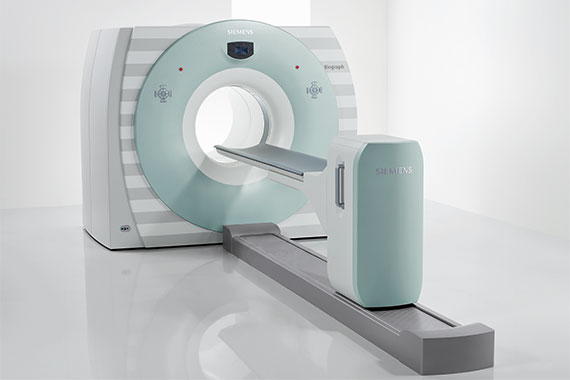
The combination of PET and CT (PET/CT) combines the functional imaging capabilities of positron emission tomography with the anatomical-morphological representation in computer tomography. Therefore, this method is particularly suitable in the care of oncological patients for assessing the extent of the disease before initiating therapy; this is especially true when it comes to determining whether and at what point a surgical intervention can improve the chances of recovery. In addition, the combined procedure documents – during and after treatment – the status of the response to the cancer therapy.
The following examinations are carried out in the Radiology Center:
- 18F-FDG: This uses a radioactively labelled sugar solution that makes it possible to visualise the energy requirements of tumours, for example, but also brain metabolism.
- Oncology: PET in combination with CT as PET/CT (imaging of tumour activity); for diagnosis, “staging” and monitoring of therapy response in many tumours.
- Neurology: metabolic function of the brain, dementia diagnostics.
These examinations are performed jointly by Kalinowski and Peloschek, specialists in radiology OG and the nuclear medicine practice, Prof. M. Hoffmann. These examinations are to be paid for privately and can be submitted to a supplementary insurance/private insurance.
- PET/CT FDG Breast
- PET/CT FDG Thyroid gland
- PET/CT FDG Prostate
- PET/CT PSMA
- PET/CT PSMA
- PET/CT radiotherapy planning chest cavity
- PET/CT radiotherapy planning
- Somatostatin receptor (SSTR-) PET/CT.
- PET/CT FDG Rectum
- PET/CT FDG Ovaries
- PET/CT FDG inguinal lymph nodes
- PET/CT FDG neck lymph nodes
- PET/CT FDG Cervix
- PET/CT FDG abdominal lymph nodes
- PET/CT FDG Axilla
- Lung cancer staging
- FET PET/CT
- F-DOPA PET/CT
- F-Cholin PET/CT

